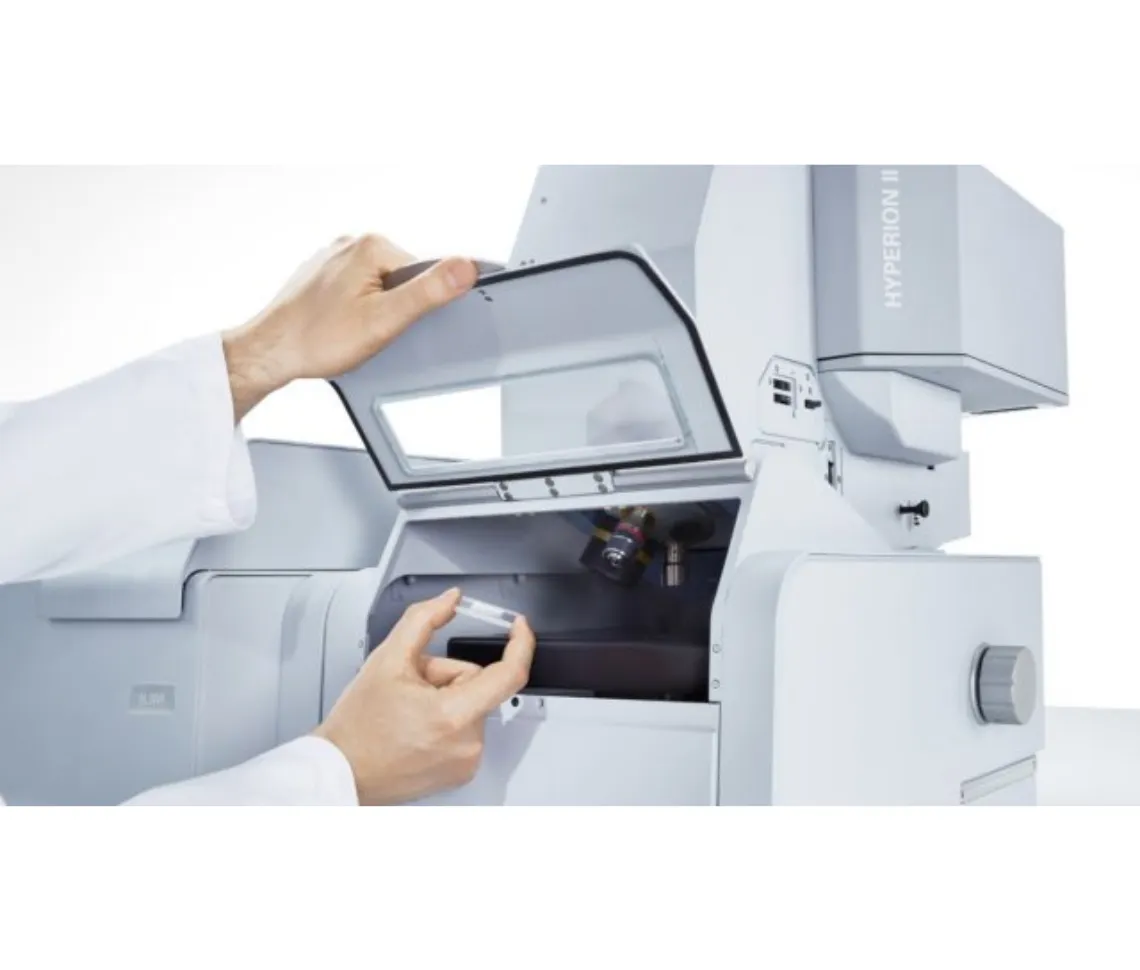
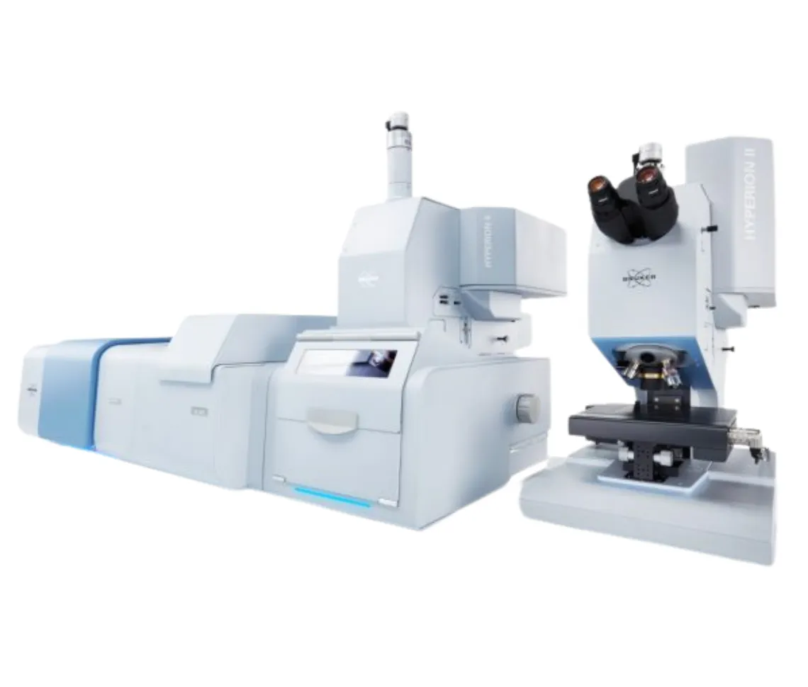
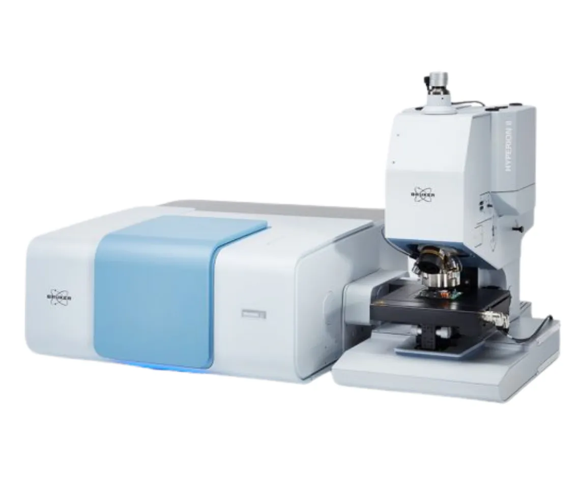
| Product Name |
HYPERION II – FT-IR Research Microscope |
| Main Purpose |
Research microscope for pioneers and innovators, flexible and configurable for complete experimental control |
| Detector Selection for µ-FT-IR |
Broad-, mid, narrow-band LN2-MCTs, Thermoelectrically cooled (TE) MCT |
| FPA Imaging Detector |
Focal-plane array detector for infrared imaging, 64 x 64 or 128 x 128 pixel |
| QCL Implementation |
Optional Laser Infrared Imaging Module (ILIM, laser class 1) |
| Objective Lens Selection |
3.5x/15x/36x IR, 20x ATR, 15x GIR, 4x/40x VIS |
| Spectral Range |
Extension from Near-Infrared (NIR) to Far-Infrared (FIR) |
| Aperture Selection |
Manual knife-edge, Automated knife-edge aperture wheel, Metal apertures for NIR |
| Accessories and Sample Stages |
Macro IR imaging accessory, Cooling/heating stage, Sample compartment |
| Visual/Optical Tools |
Darkfield illumination, Fluorescence illumination, VIS polarizers, IR polarizers |
| Perfect Image Matching |
Perfect match of spectral and visual images in any measurement mode including ATR imaging |
| FT-IR Performance |
Diffraction limited high sensitivity FT-IR microscopy and imaging using focal plane array (FPA) detector |
| FT-IR and QCL Combination |
First ever combination of FT-IR and QCL technology by optional ILIM (laser class 1) |
| IR Laser Imaging Modes |
Available in all measurement modes (ATR, Transmission, Reflectance) |
| Coherence Reduction |
Patented coherence reduction for artifact free laser imaging measurements without sensitivity or speed loss |
| Imaging Speeds |
0.1 mm² per second (FPA, full spectrum), 6.4 mm² per second (ILIM, single wavenumber) |
| TE-MCT Option |
Optional TE-MCT detector for IR microscopy with high spatial resolution and sensitivity without liquid nitrogen |
| Additional Capabilities |
Emission spectroscopy capability, Optional spectral range extensions |
| Applications |
Life science and cell imaging, Pharmaceuticals, Emissivity studies (e.g. LEDs), Failure and Root Cause Analysis, Forensics, Microplastics, Industrial R&D, Polymers and Plastics, Surface characterization, Semiconductor |
| Design Philosophy |
Complete access to instrument, experiment, samples, and parameters, Full control at any point to influence results, Precise tool that only does what user demands |
| Comparison to LUMOS II |
LUMOS II relieves user of tedious experimental details and automates measurement process, HYPERION II provides precise control and flexibility for research applications |
| Heritage |
Based on almost 20 years of HYPERION innovation in IR microscopy and imaging, All established features improved and enhanced, Sets benchmark in FT-IR microscopy and imaging |
| QCL and FT-IR Integration |
First IR microscope combining FT-IR and QCL technology in one instrument, Opens new door to life science and material research, Collect FT-IR spectrum then create QCL chemical images in seconds |
| QCL Performance |
Uncompromised QCL microscopy in state-of-the-art FT-IR microscope, Specifically developed and patented novel coherence reduction technology, Unparalleled IR laser imaging performance without digital post-processing |
| Coherence Management |
Addresses spatial coherence phenomena (fringes and speckles) that occur with QCL, Separates sample’s chemical information from physical information of scattered photons, Smart hardware design for artifact-free chemical imaging data |
| FT-IR vs QCL |
Both techniques have distinct advantages, QCL records data significantly faster at same signal to noise, QCL limited to small range of MIR, FT-IR provides universality and full spectral range, HYPERION II combines both: exceptional FT-IR imaging microscope and ambitious QCL microscope |
| Biological Tissue Analysis |
Huge potential of QCL technology for life science, Microtome section analysis with IR laser image superimposed on visual data |
| Material Science |
IR imaging for multilayer structures analysis, High-resolution ATR imaging for paint chip examination |
| Forensical Sciences |
Outstanding tool for forensic science, Fiber examination for origin evidence, Knife-edge apertures ensure optimal spectral quality |
| Drug Development |
Determining ingredients of mixtures, Pharmaceutical pellet analysis for impurities |
| Geology and Mineralogy |
IR laser imaging evaluates minerals and geochemical properties, Differentiation of oxide minerals based on reflectance properties |
| Microplastics Analysis |
FT-IR imaging as gold standard, IR laser imaging catching up, Automated microplastics analysis with particle reports and statistics |
| OPUS 9.0 – A.I.D. Feature |
Autonomous Composition Identifier (A.I.D.) – AI-driven software tool for chemical composition identification, Multi-step algorithm finds best match from reference spectral databases, Works with pure substances or complex mixtures, Automatically generates diagnostics and visual reports, High-performance results in seconds |
| OPUS 9.0 – Co-local Measurement |
Part of OPUS/OBJECT package, Seamless analysis of same region using both IR and Raman microscopy, Transfers overview images and measurement areas between LUMOS II, HYPERION II, and SENTERRA II, IR and Raman data collected from precisely same spot, Improves data correlation for heterogeneous or complex samples |
| OPUS 9.0 – Particle Identification |
Part of OPUS/OBJECT package, Combines “Find Particles” and “Cluster ID” into one workflow, Detects particles, determines dimensions, and identifies chemical makeup in one go, Simplifies analysis and accelerates decision-making |
| OPUS 8.7 – Adaptive K-means Clustering |
High performance chemical image generation, Non-supervised and autonomous determination of spectral variance, Algorithm predicts all included chemical classes by itself, Important for chemical imaging and distribution analysis, Saves valuable time |
| OPUS 8.7 – Cluster ID Function |
Identification of clusters within imaging and mapping data, Easy determination of chemical identity of classified sample components, Reliable and comprehensive statistics reports about quantity, size and identity, Leads particle and technical cleanliness analysis to new autonomous level |
| OPUS 8.7 – Updated Find Particles |
Novel particle detection method, Applied to both visual and IR image, Particle detection based on chemical images, Postrun particle determination allows determination of quantity and size from imaging or mapping results |