Description
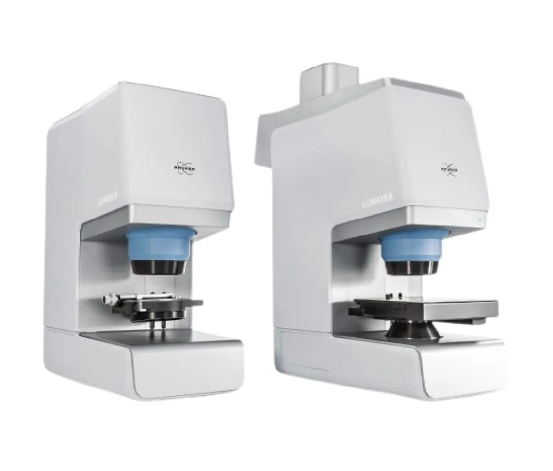
Here’s a comprehensive summary of the LUMOS II FT-IR Microscope in table format:
| Category | Details |
|---|---|
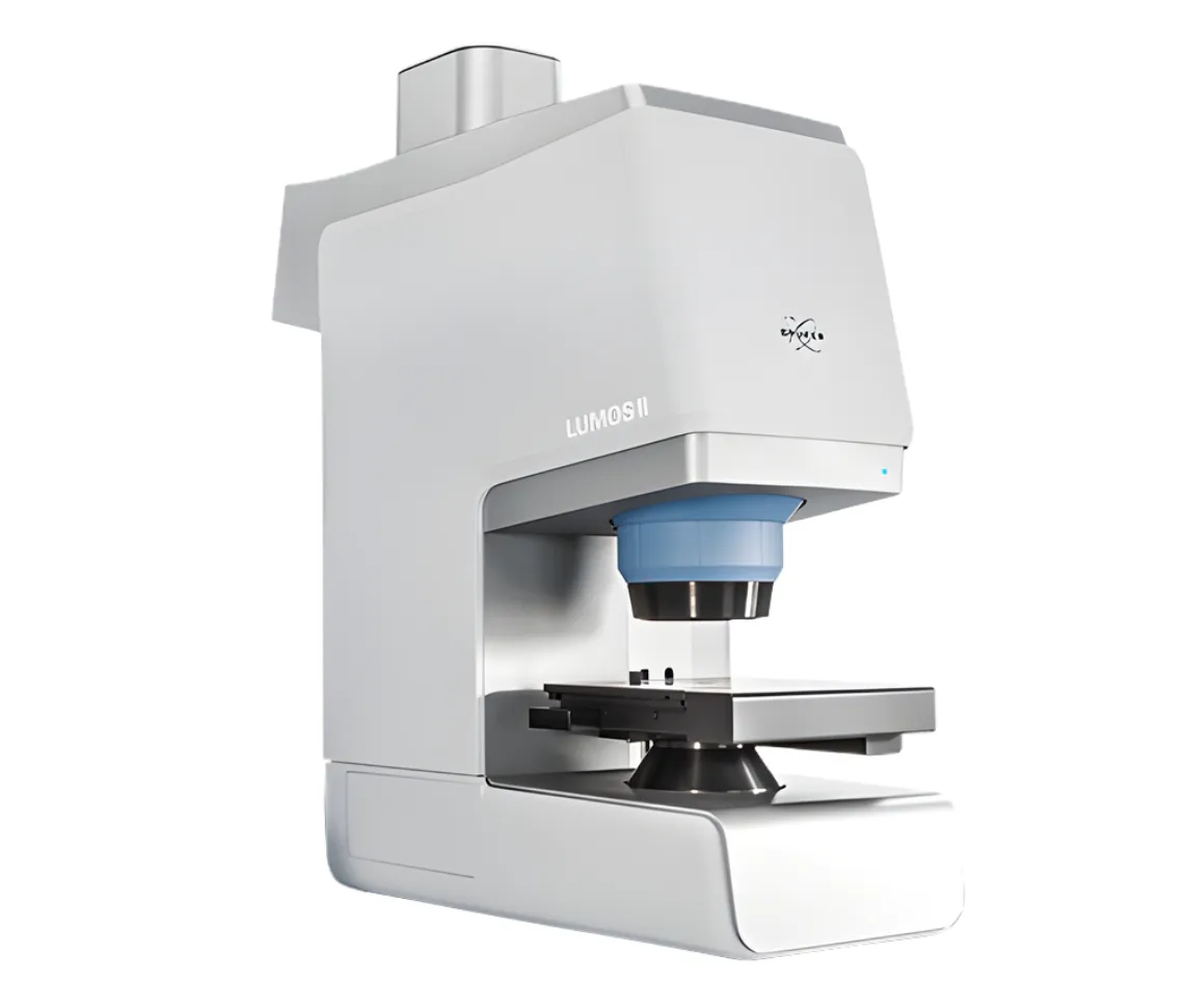
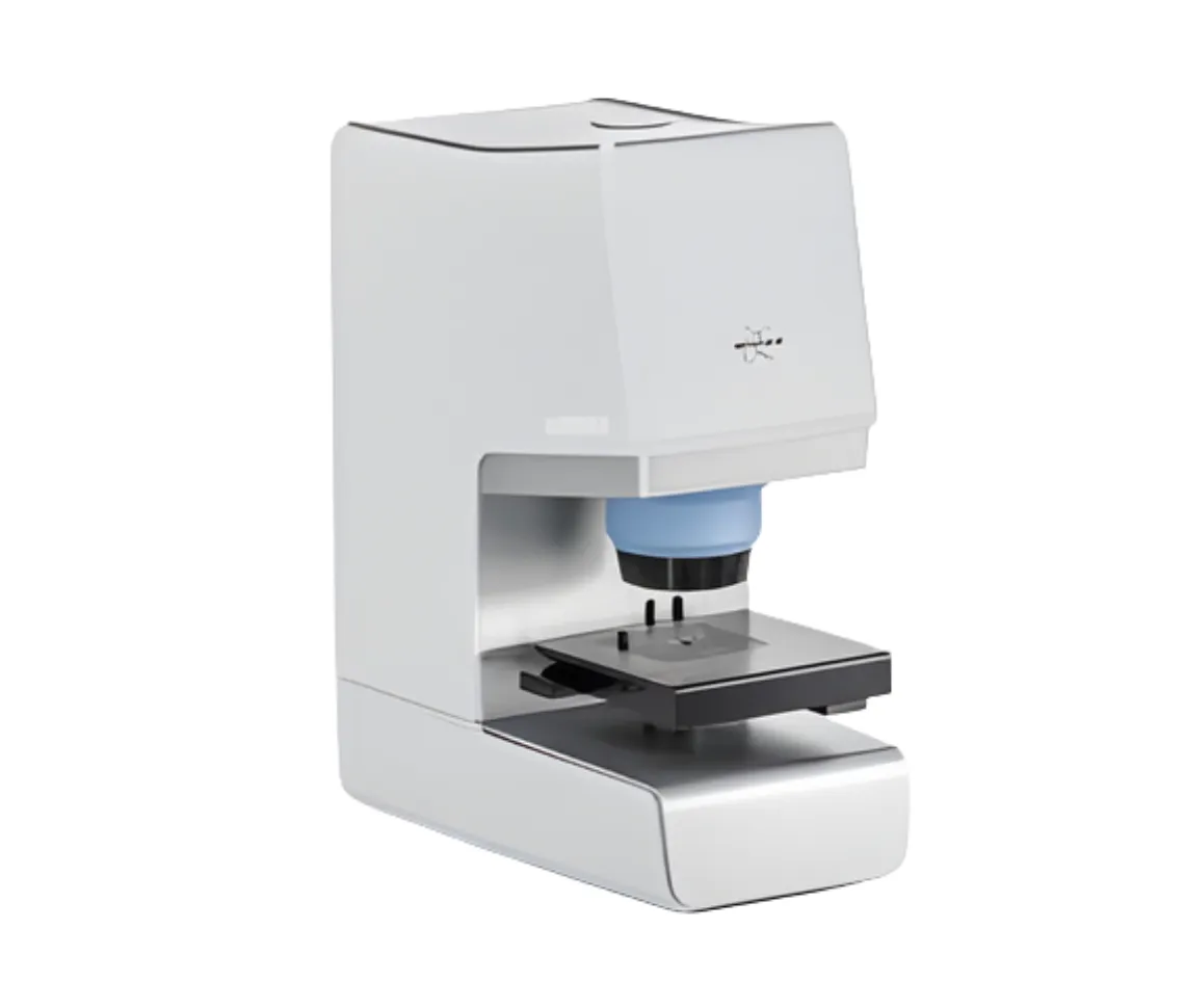
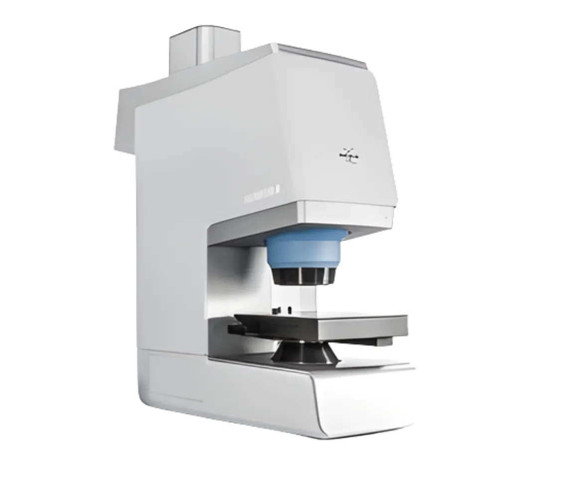
| Product Name | LUMOS II – FT-IR Microscope and Imaging System |
| Main Purpose | FT-IR microscopy, FPA imaging, and IR laser imaging powerhouse |
| FT-IR Microscopy Features | TE-MCT as standard detector, Industry leading ATR crystal design, No liquid nitrogen, no dry-air purge, LN-MCT and DTGS optionally available, Up to two single-element detectors in parallel, Upgrade to FPA imaging anytime, Analyze samples up to 40 mm in height |
| FT-IR FPA Imaging Features | Focal-plane array (FPA) imaging detector, Create FT-IR images at 1.6 mm² per second, Access 1.25 µm spatial resolution (ATR), FT-IR images at full MIR spectral range, Imaging in ATR/TRANS/REFL, Patented PermaSure+ imaging calibration, Up to two additional detectors in parallel |
| IR Laser Imaging (ILIM) | Room temperature focal-plane array detector, Create IR images at 4.5 mm² per second, IR laser images at 1,800-950 cm-1, Create chemical images in TRANS and REFL, Patented spatial coherence reduction, Application workflows (Tissue, Particle, Tablet), Open design laser class 1 instrument |
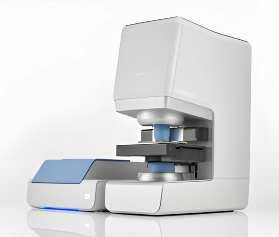
| Universal Features | Easy to use all-in-one software (LUMOS II Wizard), Inertness against high humidity using inert optical materials, AI powered evaluation routines (Adaptive Chemical Imaging), Compliance to cGMP and FDA 21 CFR part 11 with comprehensive audit trail, Complete automation and long service-life with automated knife-edge apertures, Open design with 270° access from three sides |
| Design Philosophy | Make advanced techniques available to every user regardless of skill level, FT-IR imaging faster, easier, more accurate and reliable, Tailored to user with beginners getting perfect results quickly while experts maintain total instrument control |
| Superior µ-ATR Capabilities | Unsurpassed ATR capabilities, Retractable crystal controlled by high precision piezoelectrical motors, Crystal integrated into lens, Clear view of sample while measurement takes place exactly where desired, Universal tool for failure analysis and product development, Enhanced by FPA technology |
| Technology Foundation | RockSolid™ interferometer guarantees constant performance, Modern electronics ensure mechanical precision and low energy consumption, Software monitors instrument effectiveness and ensures correct functionality |
| Application Areas | Failure analysis in plastics manufacturing, PCB failure analysis, Coating defect analysis, Adhesive layer analysis of food packaging film, Textile and fleece quality control, Fuel cell contamination investigation, Artificial leather layer checking, Plastics material research, Particle root cause analysis, Dangerous particles detection in injectable medication, Composite multi-layer polymers analysis, Geology and related sciences, Coating thickness analysis of DLC layers, Quantification in microscopic samples, Recycled plastic film characterization, Anti-corrosive coating analysis on metal surface, Multi-layer film and laminate analysis, Diamond gemstone analysis, Microplastic particles in cosmetic products, Hot chocolate powder composition investigation |
| OPUS 9.0 – A.I.D. Feature | Autonomous Composition Identifier (A.I.D.) – AI-driven software tool for identifying chemical composition, Multi-step algorithm finds best match from reference spectral databases, Works with pure substances or complex mixtures, Intelligently evaluates and recommends optimal matches, Automatically generates diagnostics and visual reports including residual spectra and hit quality graphs, High-performance results in seconds, Supports in-depth interactive analysis of alternative identifications |
| OPUS 9.0 – Co-local Measurement | Part of OPUS/OBJECT package, Enables seamless analysis of same region using both IR and Raman microscopy, Transfers overview images and measurement areas between LUMOS II, HYPERION II, and SENTERRA II instruments, Ensures IR and Raman data collected from precisely same spot, Enables complementary chemical insights, Improves data correlation and boosts confidence for heterogeneous or complex samples |
| OPUS 9.0 – Particle Identification | Part of OPUS/OBJECT package, Combines “Find Particles” and “Cluster ID” into one streamlined workflow, Detects particles within IR and Raman chemical images, Determines particle dimensions and chemical makeup in one go, Simplifies analysis and accelerates decision-making for complex samples |
| OPUS 8.8 – 3D FocusFusion | Creates pin sharp visual image even with rough or non-flat sample surfaces, Infinite “sharpness” in microscopic images helps in region of interest selection, Generates sharp visual images of samples with limited depth of field |
| OPUS 8.8 – Round Measurement Areas | FPA imaging can follow round/circular measurement areas, Measurement grid placed in round shape to measure whole particle filter, Only measures actual region of interest saving significant time during FPA measurements of round regions like filters in microplastic analysis |
| OPUS 8.7 – Adaptive K-means Clustering | High performance chemical image generation, Non-supervised and autonomous determination of spectral variance, Algorithm predicts all included chemical classes by itself, No forecasting or time consuming searching necessary, Important for chemical imaging and distribution analysis of unknown samples or small structures, Saves valuable time |
| OPUS 8.7 – Cluster ID Function | Identification of clusters within imaging and mapping data, Uses OPUS functions: spectrum search in libraries, quick compare, or identity test, Easy determination of chemical identity of classified sample components, Reliable and comprehensive statistics reports about quantity, size and identity of all analyzed structures, Leads particle and technical cleanliness analysis to new autonomous level |
| OPUS 8.7 – Updated Find Particles | Novel particle detection method, Applied to both visual and IR image, Particle detection based on chemical images measured by LUMOS II, Postrun particle determination based on chemical IR image allows determination of quantity and size from imaging or mapping results, Never miss any detail in visual or IR range |